It is almost always the first imaging study ordered to evaluate for pathologies of the thorax, although further diagnostic imaging, laboratory tests. In this article we will focus on: Elbow anatomy anatomy bones upper limb anatomy radiology schools radiology student radiologic technology medical anatomy human anatomy and physiology medical coding. It is used to evaluate the lungs, heart and chest what are the limitations of chest radiography? Chest xray is the most common examination on radiology department.
Radiology Chest Xray Normal from www.ebmconsult.com In this article we will focus on: Xray is a type of radiography and most widely used investigation. A collection of anatomy notes covering the key anatomy concepts that medical students need to learn. In fact every radiologst should be an expert in chest film reading. L the portion of the left lung that corresponds anatomically to the right middle lobe is incorporated into the left upper lobe. The interpretation of a chest film requires the understanding of basic principles. Chest xray is the most common examination on radiology department. Therefore, knowing the basics and pathologies in the ed setting is very important.
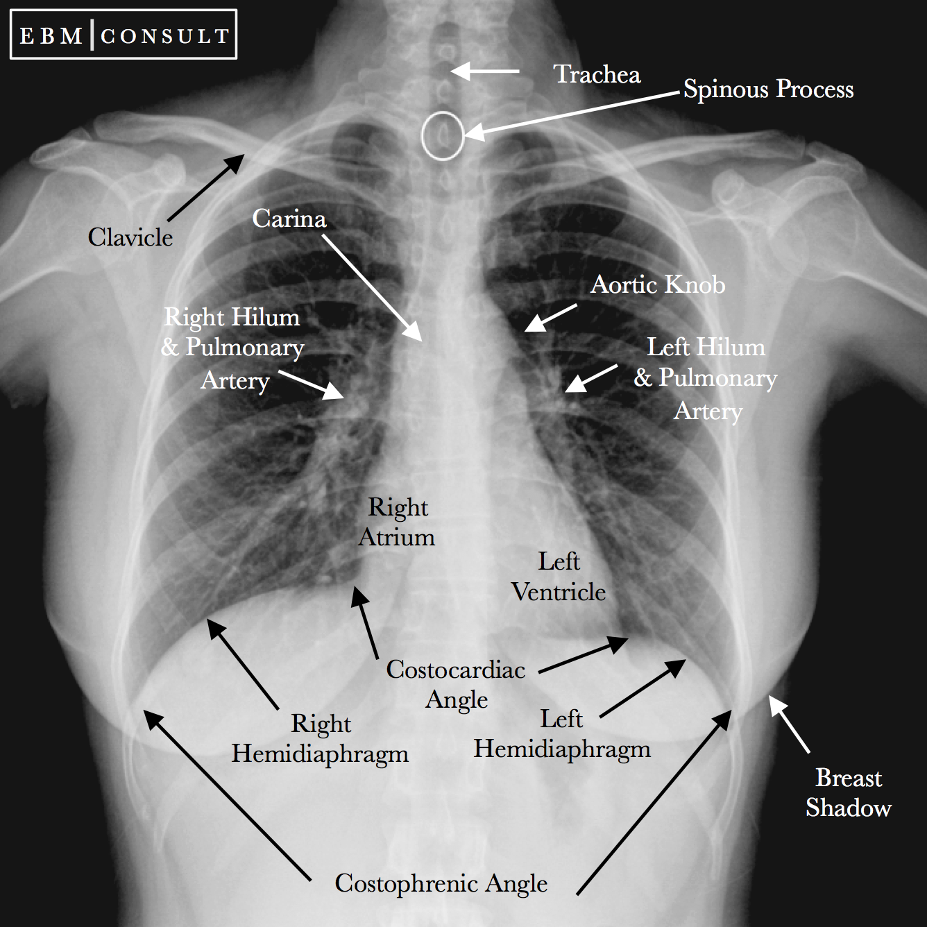
Labeled chest radiographs teaching radiologic anatomy with a level of detail appropriate for medical students.
Conclusion of living anatomy of the chest congratulations! Doctors use them to diagnose problems. Labeled chest radiographs teaching radiologic anatomy with a level of detail appropriate for medical students. The interpretation of a chest film requires the understanding of basic principles. In fact every radiologst should be an expert in chest film reading.
Chest radiographs are the most common film taken in medicine. Consolidation, interstitial, nodule/mass, and atelectasis. It is used to evaluate the lungs, heart and chest what are the limitations of chest radiography? Published 2011 by blackwell publishing ltd. Labeled chest radiographs teaching radiologic anatomy with a level of detail appropriate for medical students. Conclusion of living anatomy of the chest congratulations! Therefore, knowing the basics and pathologies in the ed setting is very important. Elbow anatomy anatomy bones upper limb anatomy radiology schools radiology student radiologic technology medical anatomy human anatomy and physiology medical coding. In fact every radiologist and pulmonary physician should be an expert in chest film reading. The interpretation of a chest film requires the understanding of basic principles. It first appears too complicated to read the chest xrays because we barely know what. This imaging method can also check how a patient is responding to specific treatments. Gillian lieberman forthe harvard 62.
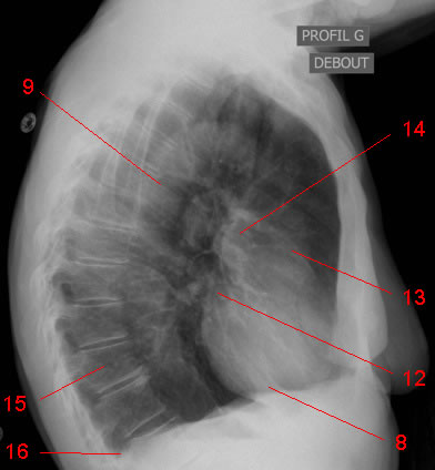
Chest X Ray W Radiology from w-radiology.com These grooves contain the neurovascular bundles that accompany each rib. Look for lung and pleural pathology. In fact every radiologist and pulmonary physician should be an expert in chest film reading. Chest xray is the most common examination on radiology department. Both lungs should be well expanded and similar in volume. L these two lobes are separated by a major fissure, identical to that seen on the right side, although often slightly more inferior in location. Common symptoms that can be diagnosed using chest. The interpretation of a chest film requires the understanding of basic principles.
Elbow anatomy anatomy bones upper limb anatomy radiology schools radiology student radiologic technology medical anatomy human anatomy and physiology medical coding.

0 Comments:
Post a Comment